


Alopecia is a condition marked by hair loss. It arises from various causes, including genetics and autoimmune disorders. Accurate diagnosis plays a vital role in treating and managing these conditions effectively. Trichoscopy in the diagnosis of Alopecia is a good idea for those who suffer from this condition.
Trichoscopy, on the other hand, is a noninvasive imaging technique. It is a reliable method for distinguishing different types of alopecia. This article examines Trichoscopy in the diagnosis of Alopecia. We will highlight its significance in diagnosing alopecia while showcasing its potential to transform hair and scalp examinations.
What is Trichoscopy in the diagnosis of Alopecia?
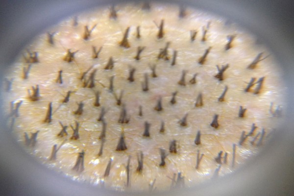
Trichoscopy, or dermoscopy of the hair and scalp, uses a specialized handheld device or video dermoscopy to magnify structures. In the diagnosis of alopecia, the trichoscopy examines the scalp and hair follicles in detail to analyze them. It can reveal patterns that help diagnose various conditions. For instance, it assists in identifying hair loss issues by providing a closer look at characteristic features.
The process involves a high-powered lens, often paired with polarized light, to enhance visibility. This magnification, ranging from 10 to 100 times, highlights specific features unique to different alopecia types. Features like yellow dots, black dots, and abnormalities in hair shafts become clearly visible, aiding accurate diagnoses.
Benefits of Trichoscopy in the diagnosis of Alopecia
By analyzing these Alopecia diagnoses with Trichoscopy and other findings, clinicians can gain valuable insights into the underlying cause of hair loss. Ultimately, this leads to a more accurate diagnosis and targeted treatment approach. The noninvasive nature of trichoscopy makes it a preferred method over traditional scalp biopsies, which can be more invasive and time-consuming. Let’s see the benefits of trichoscopy in the diagnosis of alopecia.
Treatment response
Trichoscopy is an essential tool for monitoring treatment effectiveness in individuals with hair loss. This method offers a clear view of any changes over time. As a result, doctors can detect subtle variations that indicate progress or stagnation in treatment outcomes.
By carefully analyzing these changes, trichoscopy delivers valuable insights into therapy effectiveness. This allows practitioners to make personalized adjustments to treatment plans. It also ensures they improve the results and patient satisfaction. Through this approach, trichoscopy plays a key role in achieving optimal outcomes.
Scalp biopsy

Trichoscopy is key to guiding scalp biopsies when needed. By magnifying and closely examining the scalp, it helps specialists identify areas with the most pronounced abnormalities. These may include unusual hair loss patterns, visible inflammation, or distinct skin lesions. This precise examination ensures that the selected biopsy site accurately represents the underlying condition.
Through this targeted approach, trichoscopy enhances the accuracy of the biopsy procedure. It increases the chances of collecting a tissue sample that provides critical insights for pathological analysis. Consequently, this technique significantly improves diagnostic reliability and supports effective treatment planning.
Teledermoscopy
Trichoscopy images provide a detailed view of hair and scalp conditions. It can be captured and stored digitally for further analysis. Teledermoscopy allows for easy sharing of these images with specialists across various locations.
This technology facilitates remote discussions about complex cases and enables professionals to seek second opinions. It fosters enhanced collaboration and informed decision-making among experts tackling challenging diagnostic situations.
Trichoscopy for the diagnosis of Alopecia
As we said, Trichoscopy is a valuable tool in the diagnosis of a wide array of hair and scalp disorders. This specialized technique allows practitioners to differentiate between scarring alopecia (cicatricial), where hair follicles are permanently damaged. It also helps detect non-scarring forms of hair loss.
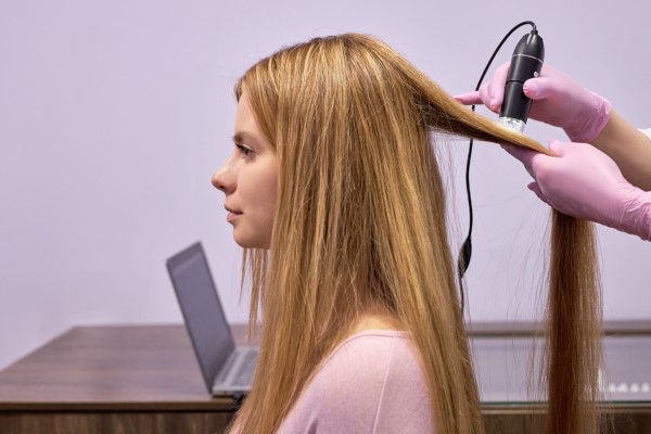
Additionally, trichoscopy enables doctors to identify the early signs of androgenetic alopecia and other types. This diagnostic approach can be instrumental in predicting the prognosis for conditions such as alopecia areata. Trichoscopy in the diagnosis of alopecia helps to find treatment options and manage patient expectations effectively.
When can I use Trichoscopy in the diagnosis of Alopecia?
Trichoscopy can be particularly useful in diagnosing challenging cases of alopecia. In some instances, distinguishing between different types of hair loss can be difficult based on clinical evaluation alone. Trichoscopy magnifies the hair and scalp structures. It allows dermatologists to identify specific trichoscopy features that aid in the diagnosis of challenging alopecia cases. Here are some scenarios where trichoscopy can be particularly helpful:
Scarring alopecia
Trichoscopy in the diagnosis of alopecia is a valuable tool for distinguishing scarring alopecia from non-scarring alopecia. These are two conditions that affect hair loss differently. In scarring alopecia, hair follicles suffer irreversible damage, leading to permanent hair loss. During the Trichoscopy in the diagnosis of alopecia, specialists can observe key indicators, such as the complete absence of follicular openings, signaling the destruction of hair-producing structures.
Additionally, white dots may appear, indicating fibrosis, or the thickening and scarring of connective tissue. Trichoscopy can also reveal areas with reduced or absent vascularity. That suggests a lack of blood supply to the affected area, further highlighting the severity of the condition.
Non-scarring alopecia
Conversely, non-scarring alopecia preserves the integrity of the hair follicles. In this case, trichoscopy typically reveals preserved follicular openings, indicating that the follicles remain intact and capable of potential regrowth.
Furthermore, normal vascularity is generally evident. It suggests a healthy blood supply to the area, which is crucial for sustaining hair growth. By comparing these distinct features, trichoscopy aids clinicians in accurately diagnosing the type of alopecia and determining the most appropriate course of treatment.
Telogen effluvium
Telogen effluvium is a medical condition marked by extensive and often sudden hair loss affecting large areas of the scalp. Trichoscopy in the diagnosis of alopecia effectively interprets the underlying causes of this hair loss. In particular, during cases of telogen effluvium, a trichoscopic analysis might reveal an increased presence of telogen hairs. These are currently in the resting phase of the hair growth cycle. This phase is crucial to understand, as it indicates a disruption in the natural hair shedding and growth process.
Furthermore, the examination may uncover the presence of short regrowing hairs and tapered hairs. That serves as a significant indicator of the hair follicles’ response to the ongoing shedding. These findings can provide essential insights into the health of the scalp and the hair growth cycle. They also help tailor effective treatment options for individuals experiencing diffuse alopecia, or Telogen effluvium.
Androgenetic alopecia
On the other hand, androgenetic alopecia, commonly known as hereditary hair loss, is characterized by distinct changes in the hair follicles. Trichoscopy in the diagnosis of alopecia can highlight the presence of miniaturized hairs, which are finer and shorter than normal.
This condition may also present signs of hair follicle miniaturization. This leads to a gradual decrease in hair density and volume over time. Understanding these patterns through trichoscopy in the diagnosis of alopecia can significantly aid in the diagnosis and management of diffuse alopecia.
Cicatricial alopecia subtypes
Trichoscopy permits the diagnosis of the different subtypes of cicatricial alopecia, including lichen planopilaris, frontal fibrosing alopecia, and discoid lupus erythematosus. Each of these conditions presents unique trichoscopic characteristics that can provide insight into their specific nature.
For instance, lichen planopilaris may display prominent perifollicular scaling and erythema. On the other hand, frontal fibrosing alopecia could show distinctive patterns of follicular loss along with changes in hair follicles. Discoid lupus erythematosus often reveals signs of follicular hyperkeratosis alongside pronounced areas of follicular dropout. These specific features help dermatologists identify and differentiate alopecias effectively. They can find the presence of perifollicular scaling and erythema, along with the degrees of follicular involvement.
Alopecia areata variants
Trichoscopy is essential in accurately diagnosing atypical variants of alopecia areata. This includes conditions like the ones we will mention above.
1. Ophiasis
Ophiasis is a variant of alopecia areata in which hair loss occurs along the periphery of the scalp, particularly on the sides and back. Trichoscopy helps diagnose this condition by revealing the absence of follicular openings and broken hair shafts in the affected areas. Recognizing these signs is crucial for differentiating opiates from other types of hair loss.
2. Sisiapho
Sisiapho presents as a band-like thinning pattern of hair, often extending across the sides of the scalp. Trichoscopic examination reveals yellow dots, which indicate sebum accumulation or follicular occlusion. On the other hand, black dots are remnants of broken hair shafts or empty hair follicles.
3. Diffuse Alopecia Areata
Diffuse alopecia areata results in even hair loss across the scalp. Trichoscopy detects this condition by showing key features such as exclamation mark hairs, which are short, broken hairs resembling exclamation points.
Identifying this specific characteristic of trichoscopy in diagnosing alopecia is crucial. It helps distinguish types of alopecia areata from other forms of hair loss. This distinction ultimately facilitates a more accurate diagnosis. As a result, it allows the development of a personalized treatment plan.
Faq’s
How does trichoscopy in diagnosing alopecia differ from a scalp biopsy?
While both trichoscopy and a scalp biopsy help diagnose alopecia, trichoscopy is a non-invasive tool that allows real-time observation of the scalp, In contrast, a biopsy requires tissue removal for pathological analysis. Trichoscopy helps guide biopsy decisions by identifying areas with the most significant abnormalities.
Is trichoscopy painful?
Not at all! Trichoscopy is a completely safe procedure. It does not involve needles or incisions, ensuring a comfortable experience for patients. Most people report feeling at ease during the examination. That is because it involves using a specialized microscope to examine the hair and scalp without causing any pain or discomfort.
Can trichoscopy detect the cause of hair loss?
Trichoscopy helps identify the type of alopecia or hair loss. Still, it does not directly identify the underlying cause. However, it can reveal patterns and characteristics that guide further testing or treatment to determine the root cause.
Conclusion
Trichoscopy in the diagnosis of Alopecia has proven to be an indispensable tool. This non-invasive technique allows dermatologists to closely examine the scalp and hair follicles, revealing crucial signs that aid in distinguishing between different types of hair loss. By identifying features such as exclamation mark hairs, yellow and black dots, and changes in follicular structure, trichoscopy enables accurate diagnosis, which is essential for effective treatment planning. Whether it’s used to monitor treatment progress, guide scalp biopsies, or identify challenging cases, trichoscopy enhances both the accuracy and efficiency of alopecia diagnosis.


Responsable – CAPIL SOLUTIONS. S.L
*Términos y condiciones
Finalidad – Dar respuesta a las solicitudes de información, y poder enviarle nuestras publicaciones, promociones de productos y/o servicios, gestionar los comentarios en el blog
Derechos Arco - Acceder, rectificar y suprimir los datos, así como otros derechos, como se explica en la información adicional.
info@capilclinic.us
Legitimación – Consentimiento
Destinatarios – No se cederán datos a terceros salvo obligación legal
Política de privacidad – Puede consultar nuestra política de privacidad aquí
*Política de privacidad